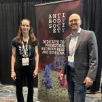
Antibody Engineering & Therapeutics commences on Thursday, December 14, 2023! Visit us at Booth #113 for a free T shirt and networking.
And don’t miss the keynote address by Dr. Heather Bax, the 2023 Huston Award recipient.
the official website of the antibody society
Antibody Engineering & Therapeutics commences on Thursday, December 14, 2023! Visit us at Booth #113 for a free T shirt and networking.
And don’t miss the keynote address by Dr. Heather Bax, the 2023 Huston Award recipient.
Photo from Adam Morse.
Summary written by Czeslaw Radziejewski, Ph.D.
Antibody Engineering & Therapeutics, held in December 2022, offered many opportunities to hear exciting and informative presentations by experts in the field, including Vaughn Smider, Ph.D., President, Applied Biomedical Science Institute, who discussed ultralong CDR H3-based knobs as the smallest antibody fragment and Jeff Allen, Ph.D. Vice President, Protein Sciences, Pelican Expression Technology, who discussed Large-scale production of knob peptides.
In 1997 Osvaldo Lopez and his colleagues [1] at the University of Nebraska analyzed transcripts encoding the variable regions of immunoglobulin heavy chains from adult and fetal bovine splenocytes. They were the first to notice the presence of long heavy chain CDR3s. The bovine CDR3s ranged in length from 13 to 28 amino acids, with the average length of CDR H3 being 21 residues in both adults and fetuses. This was longer than had been previously reported for other mammals. In a subset of bovine antibodies, CDR H3s are ultralong (50-70 AA).[2] The structure of ultralong CDR H3s was solved by Wang et al. [2] and the results demonstrated that ultralong CDR H3s all adopt similar architectures, with each composed of a long protruding beta-ribbon “stalk” and diverse disulfide-bonded “knob” (PDB: 4K3D). Up to six cysteine residues can be found in sequences of bovine CDR H3, all involved in disulfide bridges. The loops within the knob domain are thought to be involved in antigen binding. This contrasts with human antibodies where the antigen binding surface is formed from six CDR loops. Bovine CDR H3s are enormously diverse, and the diversity is generated by somatic hypermutation. [3] There is little diversity in CDR H1 and CDR H2, and cows use one light chain. Cows are not unique in having antibodies characterized by long CDR H3s. Other animals with these antibodies include zebu, yak, American and European bison.
It was previously observed that some broadly neutralizing antibodies against HIV also have longer CDR3s. Sok et al. [4] showed that immunization in cows could elicit rapid generation of neutralizing anti-HIV antibodies. Using x-ray crystallography, cryo-electron microscopy, and site-directed mutagenesis, Stanfield et al. [5] elucidated the structure of one monoclonal antibody elicited in cows by immunization with the HIV envelope trimer and showed molecular details of the knob mini-domain binding to a cryptic site on the gp120 CD4 receptor.
Knob domains are reasonably similar in size and shape to cyclotides/knottins, such as prototypic Cyclotide Kalata B1 and other disulfide-bonded peptides. Clinical applications for T cell immunotherapies are now emerging for analogs of cyclotides, for example, inhibition of the Kv1.3 channel. The Kv1.3 potassium channel is expressed abundantly on activated T cells and mediates the cellular immune responses. Sea anemone ShK cyclotide peptide was grafted into the β-ribbon ‘stalk’ of the ultralong CDR H3 scaffold of a humanized bovine IgG and showed the ability to block the Kv1.3. [6] The analog of the ShK peptide called ShK-186 or dalazatide blocks this channel, suppresses T-cell activation and is in human trials as a therapeutic for autoimmune disease.
Summary written by Alicia Chenoweth, PhD, King’s College London; Image from Ref. 7.
Antibody Engineering & Therapeutics, held in December 2022, offered many opportunities to hear exciting and informative presentations by experts in the field, including Professor Sophia Karagiannis from King’s College London, who discussed “IgE Class Antibody Immunotherapy for Solid Tumours”.
Although IgE is notorious for its role in allergic pathogenesis and anti-parasitic immune responses, there is increasing evidence that IgE may also play a role in protection against cancer. IgE deficiency is associated with increased risk of cancer, while higher total serum IgE levels may be protective against certain forms of cancer [1]. Thus, IgE biology may be of interest to cancer therapy. There are many desirable properties of IgE over the traditionally used IgG for cancer therapeutics, such as engaging powerful FceR receptors that are not shared with other classes of Ig, a very high-affinity for its high-affinity receptor FceRI which removes the need for immune complex formation allowing lower-expressing antigens to be targeted, high tissue penetration and persistence (around 1-2 weeks half-life in tissues, compared to a few days for IgG), and the lack of inhibitory receptors.
For IgE therapies, it is important to select a target which is highly expressed in tumor tissue and lowly expressed in normal tissues to enhance safety, as well as making sure that the antigen is not shed in large, multivalent formats in the circulation. Folate receptor alpha (FRa) was selected as a potential target for IgE immunotherapy, as it is overexpressed in several solid tumors, including ovarian cancer, basal breast cancer, and mesotheliomas, while also demonstrating low expression in normal tissues by transcriptomic and immunohistochemical studies [2]. An anti-FRa antibody that had already been in clinical trials and showed safety and efficacy as an IgG format was selected to engineer into an IgE format.
Summary written by Alicia Chenoweth, PhD, King’s College London
Antibody Engineering & Therapeutics, held in December 2022, offered many opportunities to hear exciting and informative presentations by experts in the field, including Dr. Hans de Haard, Chief Scientific Officer at argenx.
Dr. de Haard’s talk, Efgartigimod: A Novel FcRn Antagonist in the Treatment of Autoimmune Diseases, detailed the mechanism of action and clinical trial results of the FcRn antagonist efgartigimod. Efgartigimod is a human IgG1 Fc fragment with five “Abdeg” mutations (M252Y, S254T, T256E, H433K, N434F) to increase its affinity for FcRn at both low pH and neutral pH (1,2). It is designed to outcompete the binding of serum IgG for FcRn, leading to degradation of the unbound IgG and recycling of efgartigimod back to the surface of the cell to be released back into circulation.
Dr. de Haard discussed the findings of a recent publication in which the biochemical, structural, and in vivo properties of efgartigimod and a full-length antibody counterpart containing the same Abdeg mutations were compared (3). Crystallographic studies of FcRn in complex with the full-length antibody demonstrated that the antigen-binding fragment projects towards the membrane, leading to a potential steric clash hindering binding. This hypothesis was confirmed using a bioassay measuring receptor occupancy, showing that efgartigimod gave a better FcRn occupancy and had improved uptake compared to the full-length antibody. Furthermore, in cynomolgus monkeys, the Fc fragment gave a much faster clearance of tracer antibody and a more potent pharmacodynamic effect compared to full-length antibody. Thus, the Fc fragment was determined to be the better performing FcRn antagonist over the full-length antibody due to improved blocking of IgG recycling in vitro and the more potent PD effect in vivo.
Post written by Czeslaw Radziejewski, Ph.D.
Antibody Engineering & Therapeutics, held in December 2022, offered many opportunities to hear exciting and informative presentations by experts in the field, including Li Peng, Ph.D., who discussed “Engineering of human Sialidase Neu2 as Novel Immunotherapy for Degrading Immunosuppressive Sialoglycans to Enhance Antitumor T-Cell Immunity”.
Glycans are the most abundant structures on the cell surface. They are involved in cell communication with immune cells, and abnormal glycans can cause immune dysfunction in cancer and inflammatory diseases. Glycans typically terminate in sialic acid, but in cancer cells, sialic acid is present at a much higher abundance. The most common sialic acid in humans is N-acetylneuraminic acid, which plays a crucial role in numerous intercellular interactions, including with immune cells in the extracellular matrix, epithelial cells, and antibodies. Many studies have shown that sialoglycans are immunosuppressive and that high levels of surface sialoglycans are linked with poor outcomes in many tumor types. Hypersialylation of the surface of cancer cells makes these cells prime ligands for sialic acid-binding immunoglobulin-type lectins (Siglecs), which are found on the surface of immune cells. Once bound to sialylated glycans, Siglecs promote immunosuppressive signaling, thus conferring protection on the tumor cell. There are 15 human Siglecs. In addition, CD-28 is also known to bind sialoglycans. Most immune cells express more than one Siglec.
In her plenary lecture at the 2022 Antibody Engineering & Therapeutics conference, Professor Carolyn Bertozzi, outlined opportunities for the development of cancer treatments based on understanding the cell-surface glycome. She favored degrading sialoglycans with the enzyme sialidase to eliminate the immunosuppression promoted by Siglecs. As proof of concept, a fusion protein was created in Bertozzi’s lab using click chemistry, linking bacterial sialidase to the C-terminus of trastuzumab. The conjugate was tested in a mouse model of a trastuzumab-resistant HER2+ breast cancer model, and the results showed that the treatment essentially abrogated tumor growth. Based on these promising findings, Bertozzi cofounded Palleon Pharmaceuticals to explore sialidase-based biotherapies for cancer treatment.
At the conference, Dr. Li Peng, Chief Scientific Officer of Palleon Pharmaceuticals, presented the company’s progress in moving this concept toward the clinic. Palleon created a set of proprietary Siglec-based reagents for immunohistological hypersialylation detection and probing its role in immunotherapy resistance. Using such reagents, Palleon examined tissues from metastatic melanoma patients treated with PD1 blockade and showed that patients with a high level of sialylation fared much worse than patients with lower levels of sialoglycans. Following Bertozzi’s line of reasoning, the company pursued a strategy of using the enzymatic functionality of sialidase to remove excessive cell-surface sialylation. To translate this idea into a human therapeutic, Palleon decided to use a genetic fusion of sialidase with human Fc.