
To acknowledge and encourage the creativity of our members involved in imaging studies, The Antibody Society organized an imaging competition, open to all members.
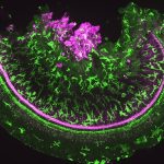
Congratulations to the 2024 Imaging Calendar Competition winner, Eva Smorodina!
 Eva Smorodina (University of Oslo and Oslo University Hospital, Norway)
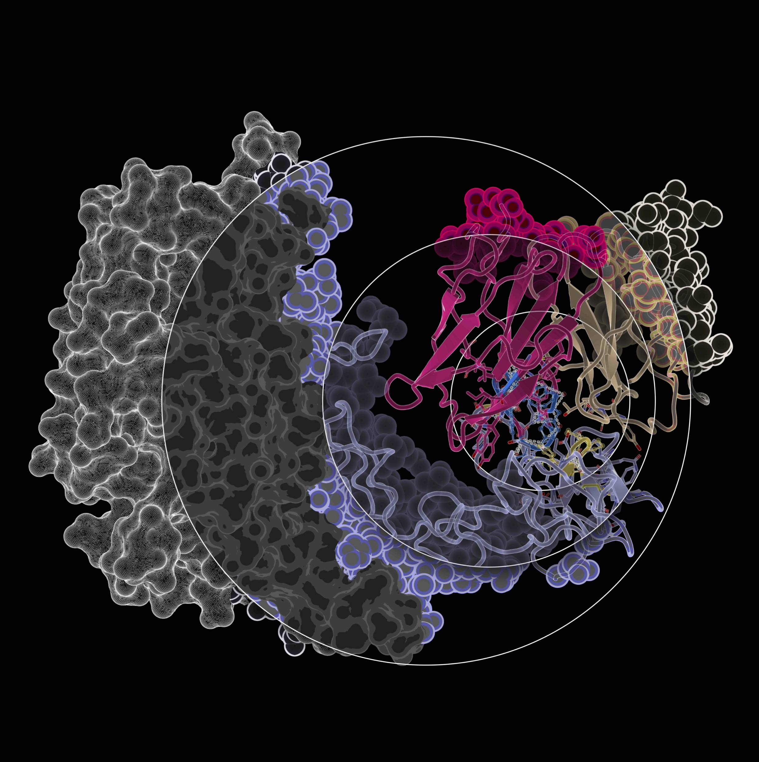
Image title: Getting closer to binding with every step
Image description: The image represents different levels of complexity surrounding antibody-antigen binding. We start from a more general understanding of the interaction kinetics with SPR, then identify the global binding site with cryo-EM, refine the region with HDX-MS to achieve peptide-level resolution, and move forward towards residue- and atom-wise resolution with computational techniques like docking and molecular dynamics. All these steps give us a better understanding of the structural rules behind antibody-antigen binding.
Antibodies used: Trastuzumab in complex with HER2
Instrument used: PyMOL, FoldX, IgFold
Congratulations also to the scientists behind the short-listed images included in The Antibody Society 2025 Calendar
-

- Eva Smorodina (University of Oslo and Oslo University Hospital, Norway): Getting closer to binding with every step.
-
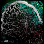
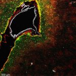
- Stefania Vilbois (University of Lausanne, Switzerland): Intravital mouse melanoma.
-
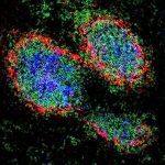
- Jessica Anania (University of Southampton, UK): Follicular Dendritic Cell Organization in Germinal Centers for Immune Memory.
-
- Andrew O’Connor (University of Southampton, UK): Inflammation in the mouse cochlea.
-

- Rodrigo García Valiente (Amsterdam UMC, The Netherlands): Clonal fireworks.
-
- Danielle Fails (Fortis Life Sciences, USA): Cells in Focus.
-
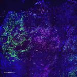
- Shaoli Lin (National Cancer Institute, NIH, USA): Therapeutic antibody targeting GPC3 in liver cancer.
-
- Jessica Anania (University of Southampton, UK): FcγRIIa resting organization.
-
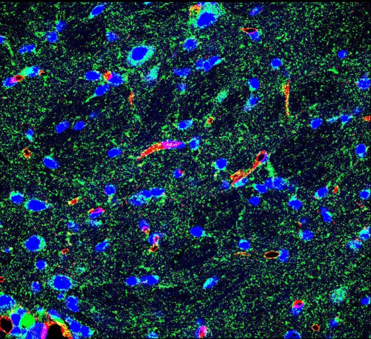
- Suzanne Buss (University of Southampton, UK): Astrocytes – helping protect our brain.
-
- Anthony Cheung (King’s College London, UK): Antibody-drug conjugates trigger microvesicle release ahead of endosomal internalization.
-
- Jessica Anania (University of Southampton, UK): Antibody kaleidoscope.
-
- Alicia Chenoweth (King’s College London, UK): Heterogeneity of melanoma.
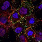
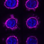
Congratulations to the 2023 Imaging Calendar Competition winner, Federica Riccio!
 Federica Riccio (King’s College London)
Image title: Surrounded
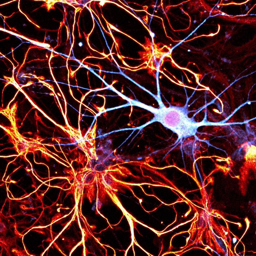
Description: A human iPSC-derived cortical neuron (MAP2 – Cyan Hot) sits happily surrounded by astrocytes (GFAP – Red Hot)
Antibodies used: rat anti-GFAP (Invitrogen 13-0300) represented in Red Hot and rabbit anti-MAP2 (Antibodies-Online ABIN1742387) represented in Cyan Hot
Instrument used: confocal laser scanning microscope Leica TCS SP8 (Leica Microsystems)
Congratulations also to the scientists behind the short-listed images included in The Antibody Society 2024 Calendar
-
- Peng Zhao (AstraZeneca): Enhanced anti-angiogenetic effect of transferrin receptor-mediated delivery of VEGF-trap in a glioblastoma mouse model
-
- Josefa Chuh (Genentech): Preclinical optimization of Ly6E-targeted ADCs for increased durability and efficacy of anti-tumor response
-
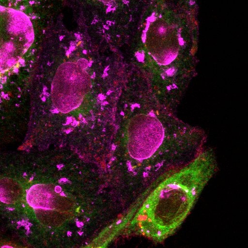
- Jessica Anania (University of Southampton): Cytoskeletal structures following FcR stimulation
-
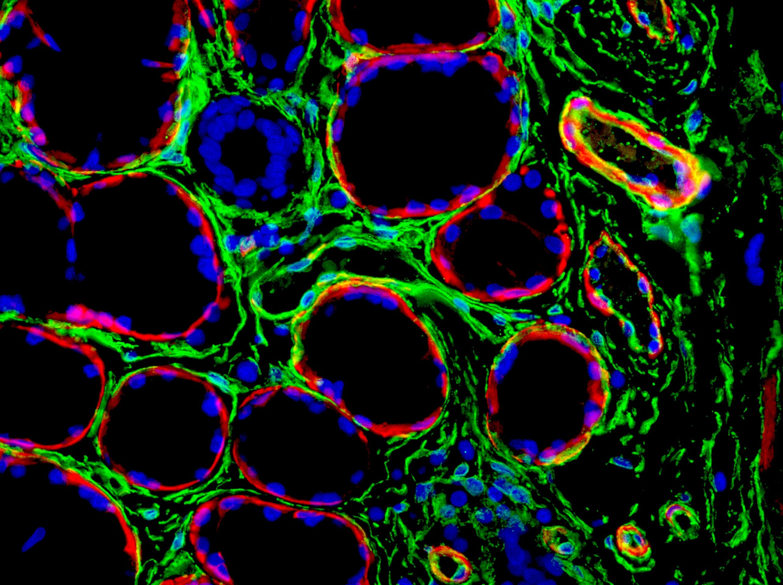
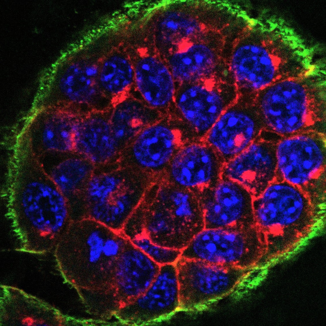
- Irene Rosa (University of Florence): Double immunofluorescence staining of human tongue, with DAPI nuclear counterstain
-

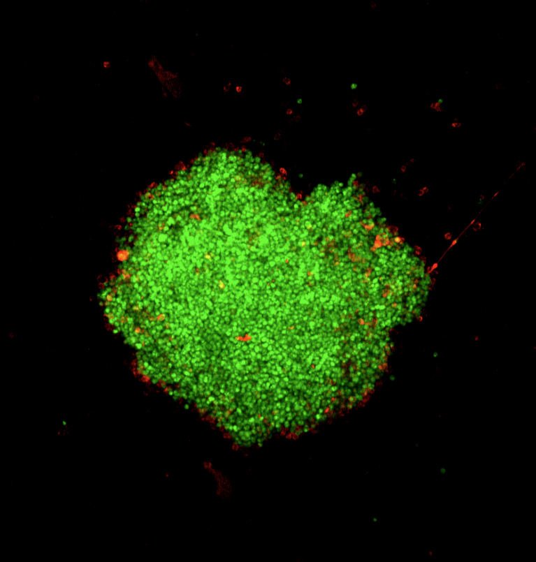
- Lorna Stewart (Fusion Antibodies): Expression bottlenecks and antibody localisation in stably transfected CHO cells with a GFP marker.
-

- Isabel Uwagboe (King’s College London): “RAGE” against the machine
-
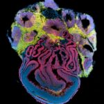
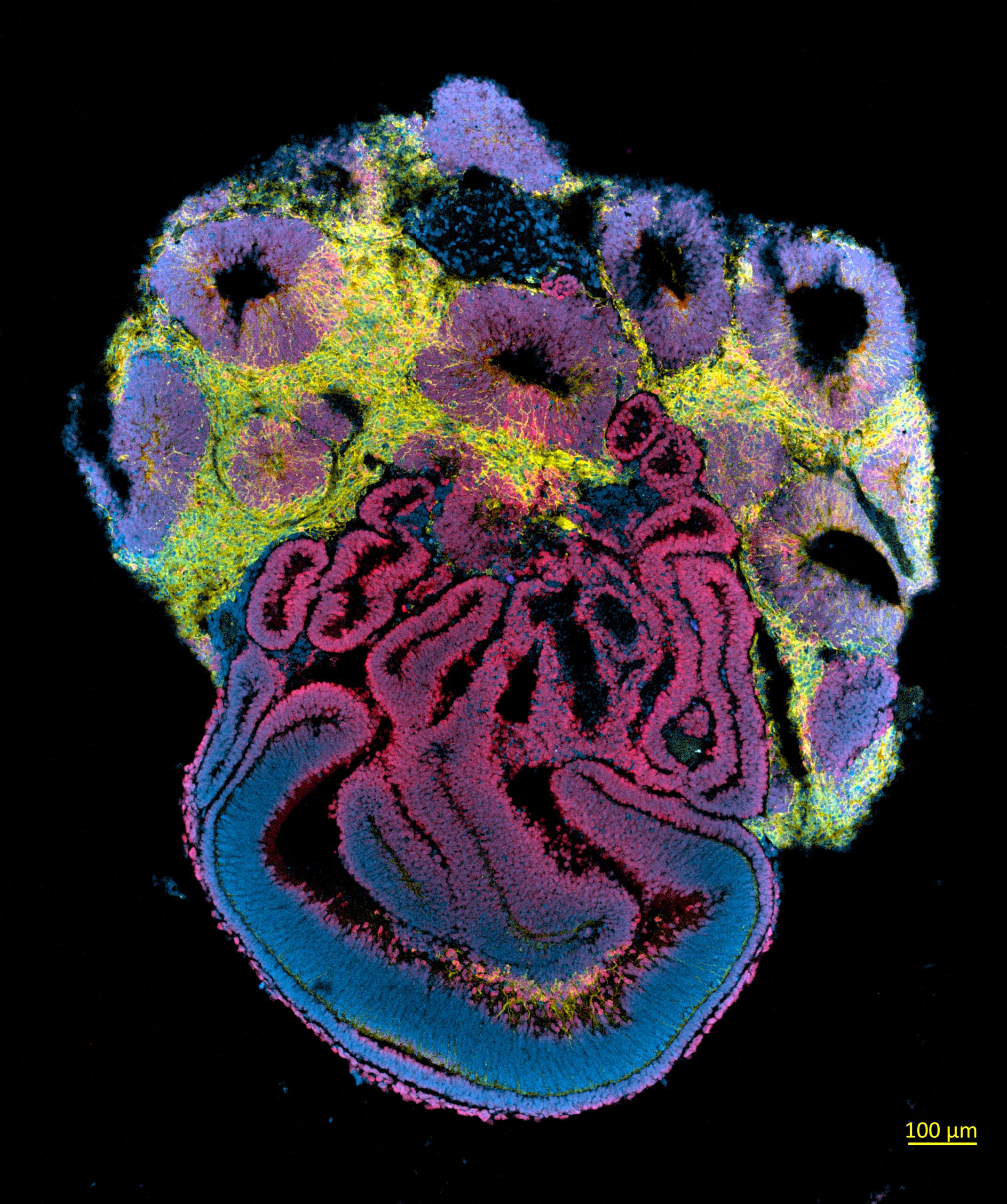
- Gabriel Emilio Herrera-Oropeza (King’s College London): Structure of an Unpatterned Cerebral Organoid
-

- Virginia Metrangolo (University of Copenhagen): Lightening up cancer cells with antibodies
-
- Sandra Lara (AstraZeneca): Antibody dependent phagocytosis of 3D tumour spheroids opsonized with Rituximab anti-CD20
-
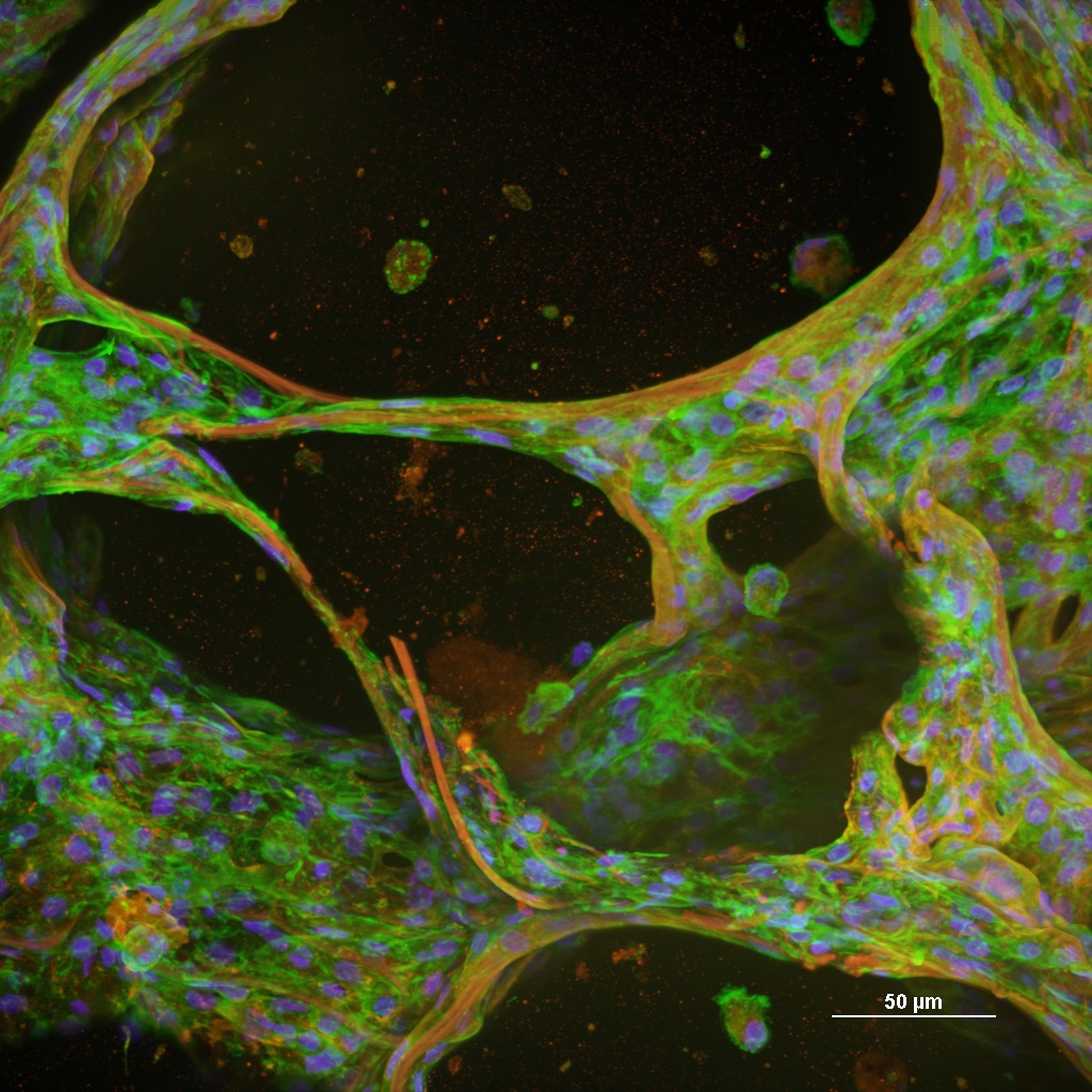
- Nathaniel Lam (GSK): Vascularised tumour-on-chip model
-
- Danielle Fails (Fortis Life Sciences): Immune profiling of axillary lymph tissues
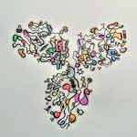
We also selected one entrant for honorable mention:
Rodrigo Garcia Valiente (Amsterdam UMC, University of Amsterdam, the Netherlands)
Image title: A(b) field of dandelions.
The image, a visual representation of a B-cell clonal repertoire using the programming language R, was not produced using microscopy and therefore didn’t meet the entry requirements. However, the Committee recognised its scientific and artistic value, and decided to give it honorable mention.