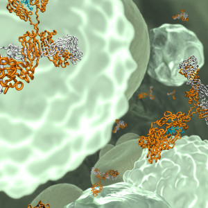
 Therapeutic antibodies have been successfully used for decades to treat various diseases. For antibodies targeting soluble antigens, however, a so-called “antibody buffering” effect, which can prolong the persistence of the target in the blood instead of clearing it, was observed. When a conventional IgG is injected into the body and binds to its corresponding antigen, the immune complexes are taken up into the cell where a certain amount of the antigen dissociates from the antibody in the endosomal compartments. The dissociated antigen is directed to the lysosomes for degradation, but the remaining amount of antigen (still bound to the IgG molecules) is recycled out of the cell by the neonatal Fc receptor (FcRn), and this can lead to an extension rather than a decrease of the antigen half-life in the bloodstream (1-5).
To overcome this buffering effect, antibodies with pH-dependent antigen binding characteristics were developed. These IgGs bind the soluble target molecules at physiological pH, but release antigen at the acidic pH in the endosomes. Antigen will then be directed into lysosomes for degradation and free antibodies will be recycled out of the cell, available for consecutive rounds of antigen binding and intracellular delivery. This method has been successfully applied to target different soluble antigens, demonstrating enhanced antigen clearance from the bloodstream compared to a conventional IgG with no pH-dependent antigen binding characteristics (6-9). Furthermore, to facilitate even more efficient antigen elimination, pH-dependent antibodies with additional modifications were generated. By increasing the antibody affinity for FcRn or FcyRIIb, soluble antigen bound to the engineered antibodies will enter the cell much more efficiently than by fluid-phase uptake. The combined effects of increased uptake and pH-dependent antigen dissociation resulted in a remarkable decrease of antigen levels following injection of engineered “sweeping” antibodies, opening possibilities for improved therapeutic applications in the future (10-13). We look forward to receiving further news about pH-dependent antibodies already in development (9, 14-15).
References:
1, Finkelman et al, J Immunol. 1993 Aug 1;151(3):1235-44.
2, O’Hear and Foote, Proc Natl Acad Sci U S A. 2005 Jan 4;102(1):40-4.
3, Phelan et al, J Immunol. 2008 Jan 1;180(1):44-8.
4, Davda and Hansen, MAbs. 2010 Sep-Oct;2(5):576-88. doi: 10.4161/mabs.2.5.12833.
5, Xiao et al, AAPS J. 2010 Dec;12(4):646-57. doi: 10.1208/s12248-010-9222-0.
6, Igawa et al, Nat Biotechnol. 2010 Nov;28(11):1203-7. doi: 10.1038/nbt.1691.
7, Chaparro-Riggers et al, J Biol Chem. 2012 Mar 30;287(14):11090-7. doi: 10.1074/jbc.M111.319764.
8, Devanaboyina et al, MAbs. 2013 Nov-Dec;5(6):851-9. doi: 10.4161/mabs.26389.
9, Fukuzawa et al, Sci Rep. 2017 Apr 24;7(1):1080. doi: 10.1038/s41598-017-01087-7.
10, Igawa et al, PLoS One. 2013 May 7;8(5):e63236. doi: 10.1371/journal.pone.0063236.
11, Iwayanagi et al, J Immunol. 2015 Oct 1;195(7):3198-205. doi: 10.4049/jimmunol.1401470.
12, Igawa et al, Immunol Rev. 2016 Mar;270(1):132-51. doi: 10.1111/imr.12392.
13, Yang et al, accepted manuscript, MAbs. 2017 Aug 8:0. doi: 10.1080/19420862.2017.1359455.
14, ALXN1210, https://clinicaltrials.gov/ct2/show/NCT02946463
15, SA237, https://clinicaltrials.gov/ct2/show/NCT02028884

